Early Decay
Spots cavities and demineralization before they become painful

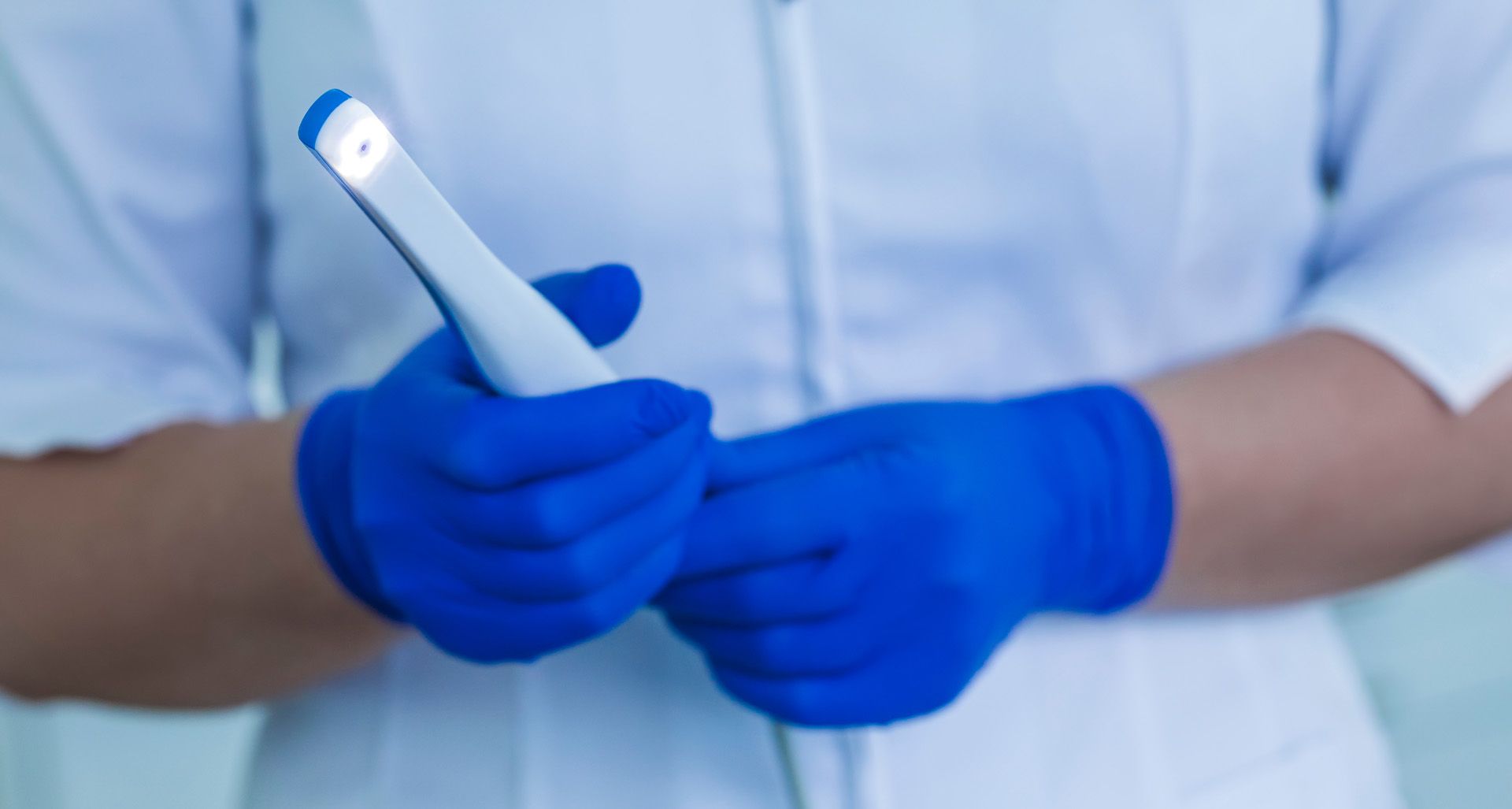
See exactly what we see, on a screen right next to your chair, so every recommendation makes sense before treatment begins.
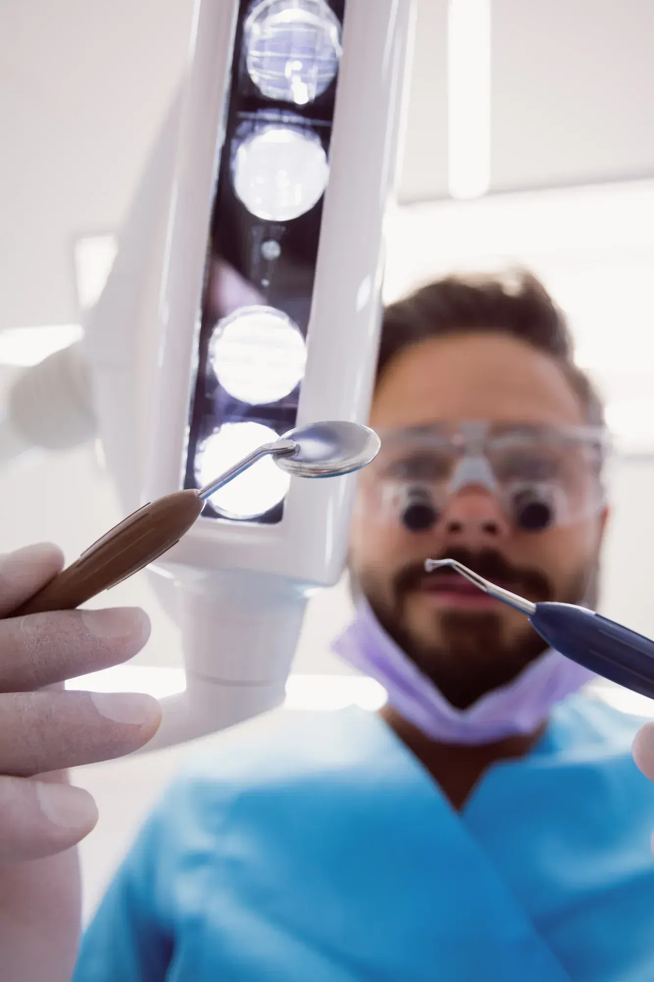
A small camera, a big difference in how you understand your own mouth.
For decades, dental visits relied on a mirror, a bright light, and a lot of trust. You sat back, opened wide, and hoped your dentist could see everything clearly enough to give you the right plan. Even with the best training, a tiny mirror tucked into the back of a mouth has limits, and patients had no way to see for themselves.
That gap is where confusion, second-guessing, and skipped treatment often begin. People say yes to fillings they do not understand or, just as often, say no because they cannot picture the problem at all.
An intra-oral camera changes that conversation. It is a slim, pen-shaped wand with a tiny lens and built-in light that fits comfortably inside your mouth. As Dr. Cook glides it across each tooth, magnified images appear on a chairside screen, sharp enough to show hairline cracks, worn fillings, and inflamed gum tissue.
You see exactly what we see. That single shift turns a one-sided exam into a real conversation about your smile, your options, and what you would like to do next.
Good to Know
A high-resolution in-mouth camera that magnifies and displays your teeth in real time
An intra-oral camera is a small, pen-shaped digital camera designed to capture detailed images from inside the mouth. Tipped with a high-resolution lens and an LED light, it connects to a chairside monitor so each image appears on screen the moment it is taken, magnifying surfaces many times beyond the unaided eye.
Tiny cracks in enamel, rough edges on old fillings, tartar along the gumline, and early gum inflammation all become easy to see and discuss. There is no radiation, no anesthesia, and no preparation - the camera simply moves around like a mirror, recording every angle in real time.
Intra-oral imaging is a quick, seamless part of your routine exam visit.
From early decay to cracked fillings, every finding becomes something you can see for yourself.
Spots cavities and demineralization before they become painful
Captures hairline fractures that mirrors often miss
Reveals worn, chipped, or leaking restorations needing repair
Documents inflammation, recession, and bleeding tissue
Shows calculus along the gumline and between teeth
Tracks grinding patterns and enamel erosion over time
Intra-oral cameras give you a clear, magnified view of your own teeth so you can understand exactly what your dentist sees and why treatment is recommended.






Every recommendation comes with a picture you can see for yourself.
Clear images make it easier to ask questions and weigh options together.
Magnification reveals problems while they are still small and inexpensive to fix.
Photos saved to your chart track changes from one cleaning to the next.
Seeing the issue removes guesswork and makes treatment choices feel certain.
Camera imaging is included with routine exams, never billed separately.
How each tool compares for accuracy, patient understanding, and recordkeeping
| Treatment | Magnification | Patient Visibility | Documentation | Hard-to-Reach Areas | Explanation Style | Radiation |
|---|---|---|---|---|---|---|
| Intra-Oral Camera | High, life-size | Live view on monitor | Photos saved to chart | Molars & back teeth | Picture-based | None |
| Dental Mirror Inspection | None | Cannot see | Verbal notes only | Limited by angle | Verbal only | None |
| Patient Hand Mirror | Naked eye only | Front teeth only | No records | Cannot reach back | Self-inspection | None |
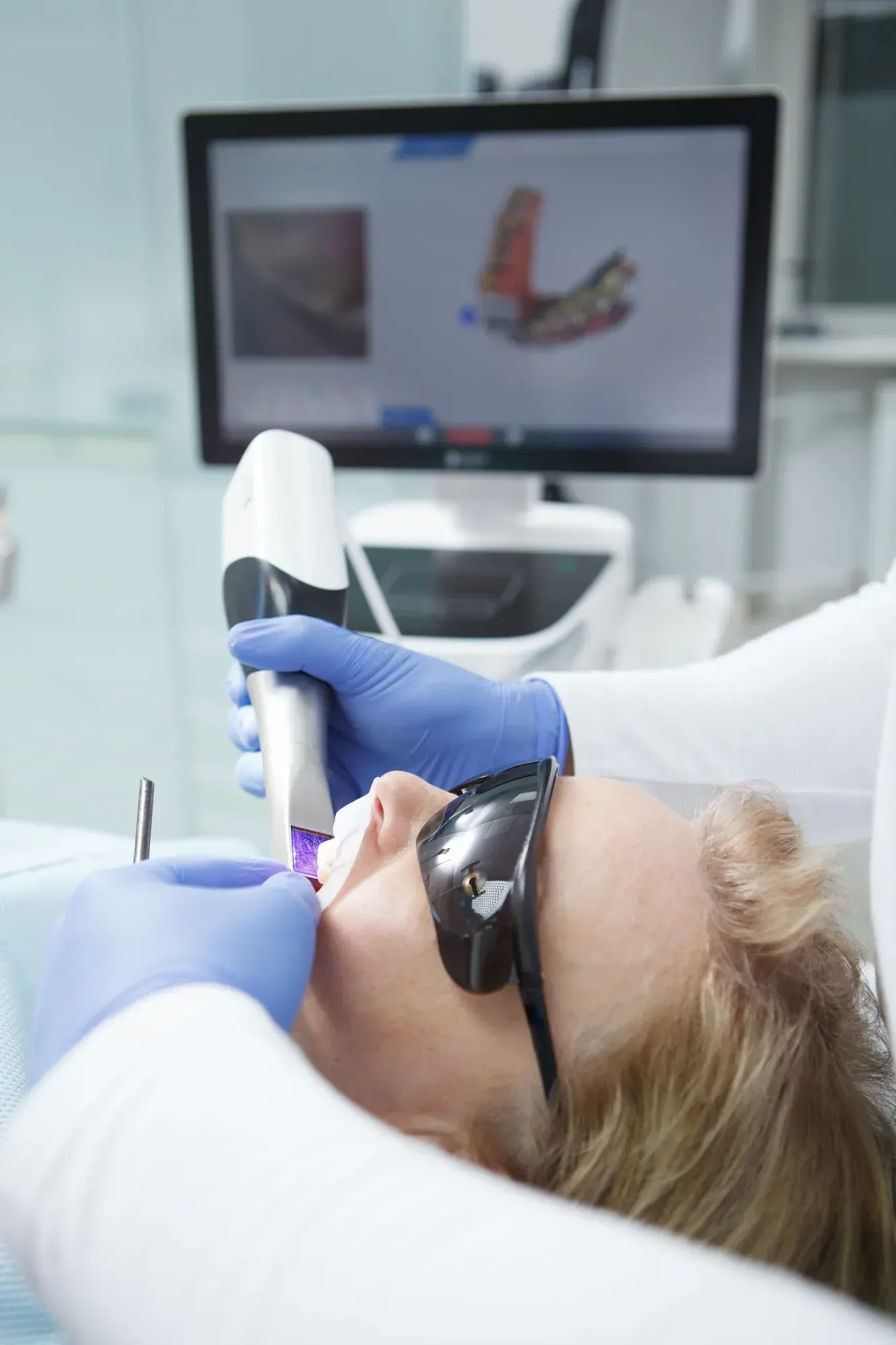
Intra-oral cameras are appropriate for nearly every dental patient, from young children visiting for their first cleaning to adults considering complex restorative work. If you have ever wished a dentist could just show you the problem instead of describing it, this technology is for you.
If any of these apply, simply tell us at check-in. We will adjust the pace, the angles, and the number of images so you remain comfortable throughout the visit.
Three effortless steps from the moment the camera wand enters your mouth to the instant images appear on the chairside screen beside you.
Dr. Cook gently positions the slim, pen-shaped camera wand inside your mouth, similar to how a small mirror would be used.
Using the camera's high-resolution lens and built-in LED light, Dr. Cook glides across each tooth and takes magnified images from several angles.
On the chairside monitor, Dr. Cook walks you through every photo, points out anything of concern, and answers questions before any treatment is recommended.
Intra-oral camera imaging is one of the safest tools in modern dentistry.
Intra-oral camera imaging is non-invasive and extremely safe - the wand uses visible LED light only, with no radiation, no medication, and no numbing. Most patients feel nothing beyond a gentle touch of the camera tip against the cheek or lip.
There are no long-term risks associated with routine intra-oral photography. Patients with a strong gag reflex may feel mild discomfort as the camera moves toward the back molars, which Dr. Cook easily manages by pacing the pass gently.
If you have a healing surgical site or a sore spot in your mouth, let Dr. Cook know before the exam so the camera can be guided carefully around that area.
Intra-oral camera imaging is included in every visit at Cook Family Dentistry - no add-on fees, no insurance surprises, just clearer, more honest care.
At Cook Family Dentistry, intra-oral camera imaging is included as a routine part of your comprehensive exam at no additional charge. There is no co-pay, no add-on fee, and no insurance code surprise - it is simply one of the tools we use to give you a clear, honest picture of your oral health.
Because intra-oral imaging is bundled into your exam, it does not require a separate insurance benefit. Our front desk team is happy to verify your coverage for routine exams and explain your specific plan before your appointment.
If any treatment is identified during your exam, we offer convenient financing options:
Our approach to transparent care along the surrounding communities.
Book NowEvery diagnosis is shown on screen before any plan is offered.
Dr. Cook combines Dawson Academy training with magnified imaging.
Kids, parents, and grandparents all benefit from clear visual care.
Serving 30A since 2010 with technology that earns your trust.
Urgent and new patient visits welcomed promptly.
Oral sedation available for anxious patients.
Complement your exam with the other diagnostic and preventive tools we offer.
View All ServicesCBCT digital X-rays in Santa Rosa Beach, FL. 3D cone-beam imaging for precise diagnosis and implant planning. Book today.
Learn moreDigital dental scanners in Santa Rosa Beach, FL. Primescan digital impressions with no messy molds. Book today.
Learn moreDental implants in Santa Rosa Beach, FL. Permanent tooth replacement that looks and feels natural. Book a consultation today.
Learn moreAll-on-4 dental implants in Santa Rosa Beach, FL. A full arch of fixed teeth on four implants. Book a consultation today.
Learn moreNot at all. The wand is smooth, slim, and only rests gently against your teeth or cheek. There is no pressure, no heat, and no need for numbing. Most patients say they barely notice it.
None. An intra-oral camera uses visible light only, the same kind of light as any digital camera. It is completely different from an X-ray and produces no radiation exposure of any kind.
No. Intra-oral camera imaging is included with your routine exam at Cook Family Dentistry. There is no separate fee, no insurance code, and no surprise bill. It is part of how we believe a thorough exam should be done.
Yes, and we encourage it. Every image appears on the chairside monitor in real time. Dr. Cook walks you through the photos, points out anything that needs attention, and answers your questions before any treatment is recommended.
Yes. Each image is stored securely in your electronic dental chart. That way, at your next cleaning, we can compare new photos to older ones and watch for subtle changes over time, which helps us catch problems earlier.
Absolutely. Kids often enjoy seeing their own teeth on the monitor, and it becomes a great teaching moment about brushing, flossing, and cavity prevention. We work at a pace each child is comfortable with.